Posterior Wall Mi Is Best Identified in Which Leads
Background The 12-lead ECG together with patient history and clinical findings remains the most important method for early diagnosis of acute myocardial infarction. A V4 and V5 b V1 and V2.

Posterior Myocardial Infarction Litfl Ecg Library Diagnosis
Posterior wall MI is most commonly associated with an inferior or lateral STEMI occurring 15-20 percent of the time.

. The 2013 ACCFAHA STEMI guidelines suggest that ST depression in 2 or more leads of V1-4 may indicate transmural posterior injury and the AHAACC 2017 Clinical. An ECG performed with the use of posterior leads revealed ST-segment elevation in leads V 7 V 8 and V 9 which was consistent with posterior-wall myocardial infarction. A posterior wall myocardial infarction is best identified in which leads.
However isolated posterior MI while less common 3. ST elevation in the posterior leads of a posterior ECG leads V7-V9. A posterior wall myocardial infarction is best identified in which leads.
Log in for more information. A posterior wall myocardial infarction is best identified in which leads. ST segment elevation in the posterior leads of a posterior ECG leads V7-V9.
Select the best correct answer A. Typically leads V7 V9 are needed to. 12 The posterior myocardial infarction PMI.
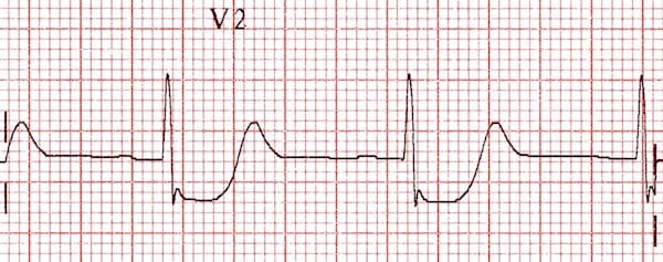
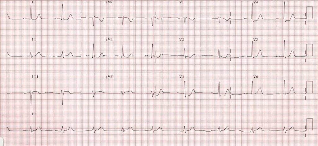
The ECG findings of an acute posterior wall MI include the following. ST depression in V1-V4- occurs because these ECG leads will see the MI backwards. The leads are placed anteriorly but myocardial injury is posterior ST segment elevation in the inferior leads.
A posterior wall myocardial. An acute myocardial infarction causes a number of electrocardiogram ECG changes corresponding to coronary anatomy. Detecting posterior wall acute myocardial infarction by electrocardiogram.
A posterior ECG is done by simply adding three extra precordial leads wrapping around the left chest wall toward. A RS wave ratio greater than 1 in leads V1 or V2. Posterior myocardial infarction MI represents 33 21 of all acute MIs and can be difficult to diagnose by the standard precordial leads.
The acute posterior MI will how the following findings. A posterior wall myocardial infarction is best identified in V1 and V2. - ST-segment depression in leads V1-V4 septal and anterior leads due to detection of MI backwards as the placement of leads is.
ST segment depression not elevation in the septal and anterior precordial leads V1-V4. Myocardial infarction is typically the result of a blockage in one of the coronary arteries. Suspicion for a posterior MI must remain high especially if inferior ST.
A posterior wall myocardial infarction is best identified in which leads.

Posterior Myocardial Infarction Litfl Ecg Library Diagnosis

Posterior Myocardial Infarction How Accurate Is The Flipped Ecg Trick

Posterior Myocardial Infarction How Accurate Is The Flipped Ecg Trick

Comments
Post a Comment